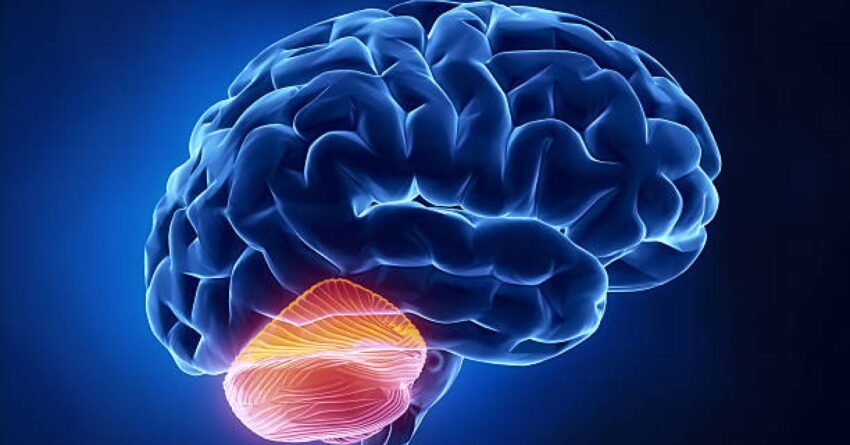
Fear is a fundamental emotion necessary for survival. We experience fear in several ways that have helped humans stay alive, but it can go beyond what is appropriate for survival. Persistent fear and anxiety are symptoms of posttraumatic stress disorder and general anxiety disorder, affecting about 8 percent of American women and 4 percent of American men. Where does this fear come from and where is it located in the brain? New research has begun to tell us that fear exists well below the conscious level in the cerebellum and is an important component of what was previously thought to be exclusively responsible for internal motor functions. New experiments examine this part of the brain with the ultimate hope that there may be improved treatments for anxiety and fear disorders.
The Role of the Cerebellum
The cerebellum, located at the back of the brain and intimately connected to the brainstem, has various motor and cognitive functions. It is well known for its role in maintaining balance, coordinating fine movements, and learning new motor skills. More recently, however, new research has demonstrated that the cerebellum contributes to certain cognitive abilities beyond motor control, such as attention, emotional processing, decision-making, and procedural learning.
Recent studies have observed the cerebellum’s role, particularly in regulating fear. Fear is necessary for survival and humans learn fear responses through exposure. The cerebellum has been shown to contribute to learned fear responses, also known as conditioned fear responses. This is when a fear of a previously neutral stimulus (for example a tone, light, or context) is developed through a learning process like classical conditioning. The neutral stimulus is consistently paired with a threatening stimulus (like an electric shock or loud noise) creating an association whereby what was once neutral becomes frightening. For example, a tone repeatedly followed by an electric shock becomes frightening, regardless of whether or not the shock follows.
Functional brain imaging shows the involvement of the cerebellum in fear conditioning in humans but does not determine the exact role or even necessity of the cerebellum on fear. One way to assess the impact of a certain brain region on behavior is to study people who have experienced brain damage or brain lesions. A recent article was published in eNeuro describing a study conducted by researchers at the Department of Neurology and Center for Translational Neuro- and Behavioral Sciences at Essen University Hospital and the Department of Behavioral Neuroscience at Ruhr University Bochum in Germany. They investigated whether patients with cerebellar cortical degeneration have alterations in the acquisition and extinction of learned fear responses.
The Cerebellum and Fear
The study included 20 cerebellar patients and 21 controls. The cerebellar patients had ataxia, a condition of poor muscle control usually caused by damage to the cerebellum. Various conditions can lead to ataxia including genetic conditions, stroke, tumors, multiple sclerosis, and degenerative diseases. All participants were given an electric shock when presented with a blue, red, or yellow light. The experiment was split into different phases: habituation, fear acquisition training, and extinction training on the first day and recall phase on the second day. Electrocardiograms and skin conductance responses were recorded using magnetic resonance imaging and electrocardiogram devices during the experiment. The participants also filled out questionnaires rating their fear.
For comparison, the study also examined fear conditioning in mice with cerebellar cortical degeneration. The mice were placed in a fear conditioning chamber where they heard a tone followed by a shock. The mice were brought to the fear extinction chamber for three consecutive days following the fear acquisition to ensure the fear was completely gone.
Study Results and Implications
Patients with cerebellar cortical degeneration showed mild deficits in fear learning compared with controls. They showed slower acquisition of fear associations and delayed consolidation of learned fear. Their extinction learning was slower, with indications of incomplete consolidation of fear associations. They required more time to recognize the associations between conditioned and unconditioned stimuli, indicating deficits in working memory processes. Behavioral abnormalities were mild compared to deficits observed in motor associative learning tasks. The researchers note that fear conditioning involves an extended neural network, including the amygdala, potentially compensating for cerebellar deficits.
The implications of the study are significant in a few ways. Primarily, the study sheds light on the involvement of the cerebellum in fear learning processes, which has been less understood compared to the role of the cerebellum in motor control. The study highlights the cerebellum’s role in cognitive and emotional processes. Additionally, the study has clinical implications for patient care and treatment development. By recognizing that patients with cerebellar cortical degeneration may exhibit deficits in fear learning, healthcare professionals can provide tailored interventions. There is also the potential to develop novel treatments or rehabilitation strategies targeting specific brain regions or pathways involved in fear conditioning, giving hope to those who may suffer from anxiety or stress disorders.
